The Biology Department Imaging Facility serves both teaching and research needs for the Biology Department as well as other departments within the College of Science and Technology, and Temple University as a whole. We work in collaboration with other resources in the college, such as the Materials Research Facility , to provide a rich array of micro- and nano- resources. Major components of the facility include:
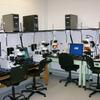
A general purpose light microscope laboratory with 10 light microscopes (Nikon, Olympus) equipped for brightfield, darkfield, phase contrast, fluorescence and differential interference microscopy. Each is coupled to a digital image capture system with appropriate analytical software.
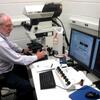
A Leica Laser Scanning Confocal Microscope is supplied with three lasers and unique spectral detection, permitting fluorescence and spectral analysis for biological and non-biological materials. Precise control of depth of penetration depth allows the formation of three-dimensional images.
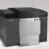
An Agilent 8500 field emission scanning electron microscope (FE-SEM) that offers resolution of up to 10 nm, and operating voltages from 0.5 to 2kV, with detection of backscatter and secondary electrons. These capabilities permit nanoscale features to be observed on a wide variety of biological and nanostructured materials, including cells, tissues, polymers, thin films, biomaterials, and other energy-sensitive samples on any substrate, even glass.